
Not every mole that worries you needs the same level of scrutiny — and not every check needs the same tools. Understanding the difference between dermoscopy and mole mapping can help you make sense of what actually happens during a skin examination, and why.
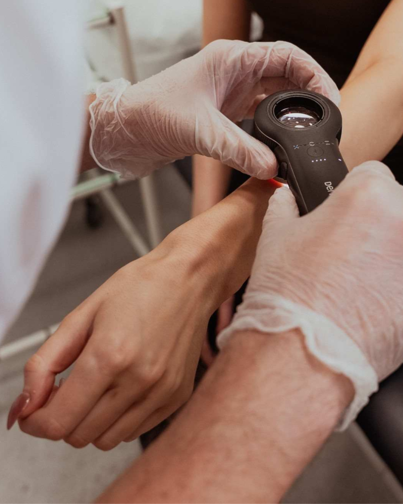
If you’ve ever had a mole checked, you’ve probably had a clinician look at it with a small handheld device pressed gently against your skin. That’s a dermatoscope, and the technique is called dermoscopy — a cornerstone of modern skin cancer screening that most patients have experienced without necessarily knowing what it’s called or why it matters.
What Is Dermoscopy?
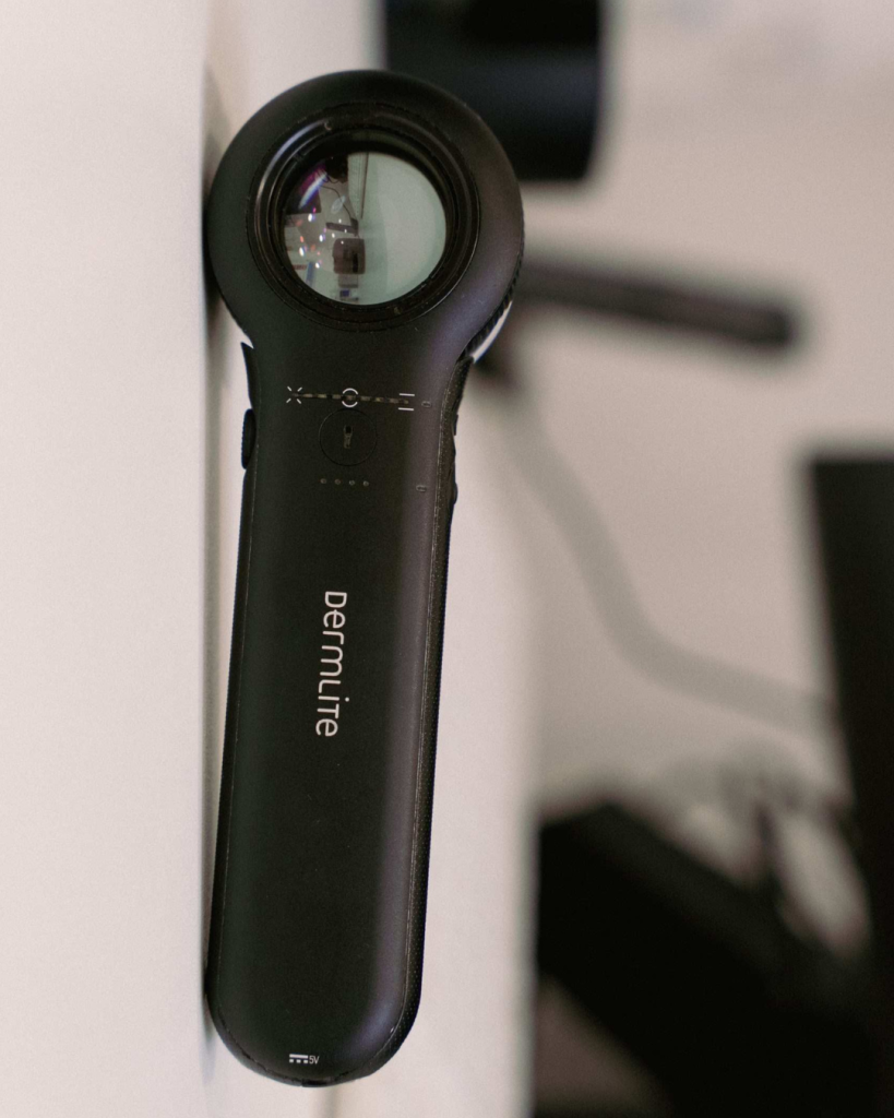
Dermoscopy is a non-invasive skin examination technique that lets clinicians see far more detail than the naked eye ever could. The dermatoscope combines two things:
- Magnification — typically around 10x
- Polarised or non-polarised light — which cuts through surface glare and reflection
Together, these let a clinician see structures just below the skin’s surface — pigment patterns, blood vessels, and skin architecture — that are otherwise invisible.
What Is It Used For?
Dermoscopy is used to evaluate:
- Moles and pigmented spots
- Suspicious lesions that could be melanoma
- Other skin cancers, including basal cell carcinoma and squamous cell carcinoma
- Hair and scalp conditions
- Nail abnormalities
- Some inflammatory or infectious skin conditions
Why It Matters
Compared with an unaided visual check, dermoscopy improves a clinician’s ability to judge whether a lesion looks benign or needs monitoring, biopsy, or removal — particularly when melanoma is a possibility.
It also cuts both ways: better cancer detection, and fewer unnecessary biopsies and excisions of harmless moles. That combination — catching more of what matters while removing less of what doesn’t — is why dermoscopy is now considered a standard part of dermatology and skin cancer screening, especially for anyone presenting with pigmented lesions or at increased melanoma risk.
What Is Mole Mapping?
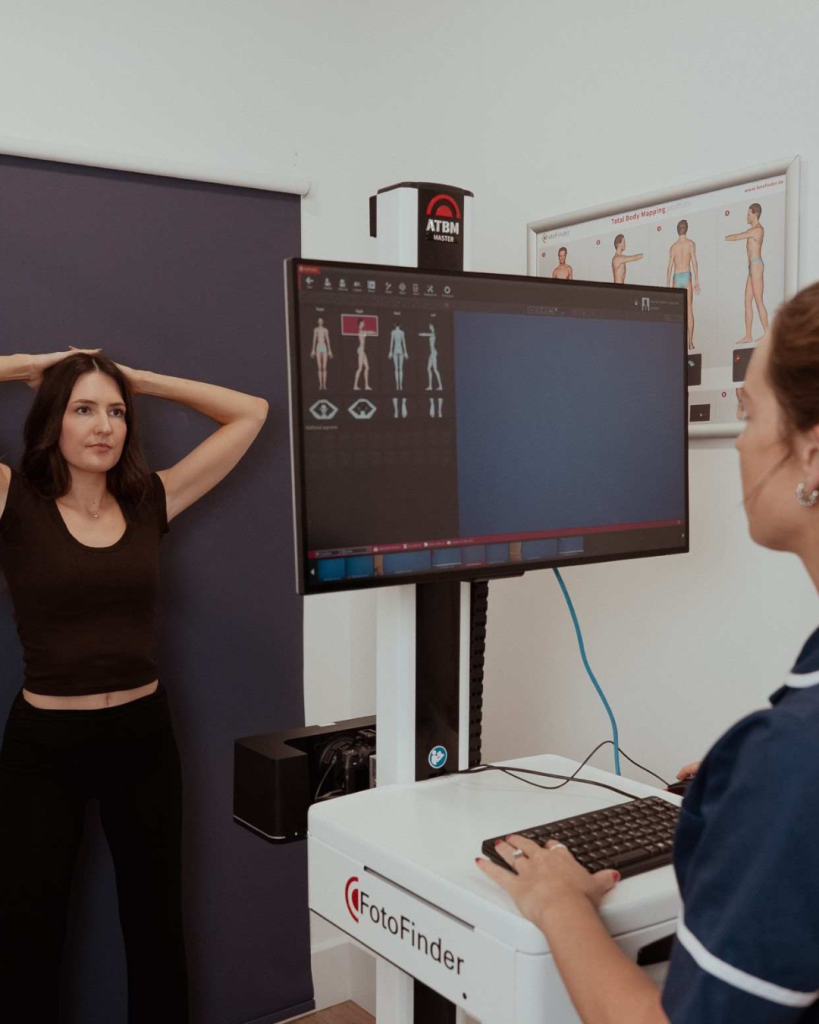
Mole mapping takes this a step further, but for a specific purpose: tracking moles over time rather than assessing them in a single visit. It combines:
- Total body photography — standardised photographs that create a baseline record of the skin
- Digital dermoscopy — magnified images of individual moles to record their structure
At follow-up appointments, these images are compared against the baseline to spot new lesions or changes in existing ones — a method built around early detection of melanoma through change over time, rather than a single snapshot.
Why Mole Mapping Isn’t for Everyone
Here’s the part that often gets missed: mole mapping — sometimes called total body photography with sequential digital dermoscopic imaging — isn’t automatically the right tool for every patient.
It’s most valuable for people at increased melanoma risk, including those with a personal or family history of melanoma, multiple atypical naevi, dysplastic naevus syndrome, or a high total mole count. The International Dermoscopy Society recommends digital monitoring primarily for these higher-risk groups, where serial imaging can pick up the kind of subtle changes that signal early melanoma (Argenziano et al., 2022).
For lower-risk individuals, a thorough skin examination supported by dermoscopy is usually enough. Routine mole mapping in these cases can add cost, extra follow-up appointments, and patient anxiety without a clear clinical upside.
So the two techniques sit alongside each other rather than in competition: dermoscopy as the core of skin lesion screening for most patients, and mole mapping reserved for those whose risk profile means long-term digital surveillance will genuinely pay off.
Which Check Is Right for You?
“Dermoscopy is part of every skin check we do — it’s a fundamental tool. But mole mapping is a different service with a different purpose, and we’re careful to recommend it only where the evidence supports it. Patients with a strong personal or family history of melanoma, or a high number of atypical moles, get real value from sequential imaging. For everyone else, a comprehensive dermoscopy-led skin check is the right level of care.”
— Dr Tim Cunliffe, Medical Director, The MOLE Clinic
At The MOLE Clinic, our dermatology-trained clinicians use dermoscopy as standard in every skin assessment, and can advise whether mole mapping is appropriate for your individual risk profile. Appointments are available across our UK clinics, with no GP referral needed. Book a Full Body Skin Check or Mole Mapping appointment today.
Early detection saves lives. Don’t leave it until something looks wrong.
The MOLE Clinic is a UK-based private skin cancer screening and dermatology provider. We offer mole checks, full-body skin examinations, and specialist dermatology services at clinics across the UK.
Reference
Argenziano, G., Badertscher, N., Cerroni, L. et al. (2022) ‘Consensus recommendations for the use of non-invasive melanoma imaging in patients at increased risk’, Journal of the European Academy of Dermatology and Venereology, 36(10), pp. 1663–1678.